A 30-million-cell atlas of the human brain—with limits
A photorealistic 3D render of 30 million brain cells, with volumetric lighting, centered in the frame with minimal negative space, neutral editorial📷 Photo by Tech&Space
- ★30 million cells from nearly 200 studies mapped
- ★Focus on early cognition and neurodevelopmental conditions
- ★No direct patient impact yet—research-stage only
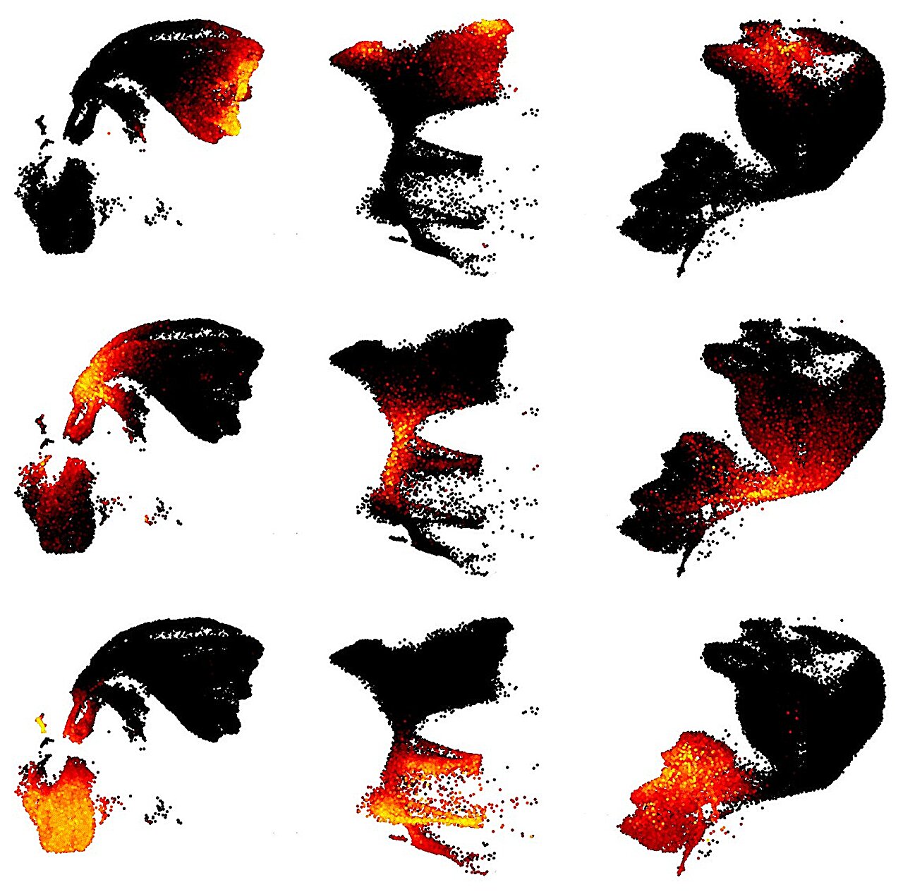
The human brain’s molecular architecture is now clearer than ever, thanks to a new atlas combining data from 30 million cells and nearly 200 studies. Led by investigators at Johns Hopkins Medicine, the project aims to decode how early brain development influences cognition and neurodevelopmental disorders—conditions like autism, schizophrenia, and intellectual disabilities that often emerge in childhood but reshape lives for decades.
This isn’t a clinical tool yet. It’s a research-stage resource, designed to help scientists identify cellular patterns linked to disease. The scale is unprecedented: previous atlases relied on far smaller datasets, often from single studies. Here, the team harmonized disparate data sources, accounting for variations in tissue sampling, sequencing methods, and developmental stages. That’s both the strength and the challenge. As co-author Dr. Seth Ament noted in the announcement, 'We’re not just adding data—we’re trying to standardize how we interpret it.'
The atlas focuses on prenatal to early postnatal development, a critical window where disruptions can have lifelong effects. But its clinical relevance today is indirect. No new therapies, diagnostics, or guidelines emerge from this work—only a foundation for future hypotheses.

The most detailed molecular map of the brain to date—still years from clinical use📷 Photo by Tech&Space
The most detailed molecular map of the brain to date—still years from clinical use
For all its ambition, the atlas has inevitable gaps. The 30 million cells represent a fraction of the brain’s estimated 86 billion neurons, and the studies included vary in quality and scope. Some datasets rely on post-mortem tissue, which may not fully reflect living brain dynamics. Others use single-cell RNA sequencing, a powerful but still evolving technique. The team mitigated these issues with computational normalization, but experts warn that such harmonization can introduce its own biases.
What the atlas does offer is a shared reference point. Researchers studying, say, how maternal infection affects fetal brain development can now cross-reference their findings against a standardized molecular baseline. That could accelerate the pace of discovery—but it won’t replace the need for targeted, mechanistic studies. The NIH’s Brain Initiative, which partially funded this work, has long emphasized that atlases are tools, not solutions.
Regulatory agencies aren’t yet involved. This is pre-clinical research, far removed from the FDA’s purview. The next step? Validating the atlas’s predictions in animal models or human cohort studies—work that could take years. In the meantime, the data is publicly available, a deliberate choice to foster collaboration. But as with all big-data projects, the real test will be whether the insights hold up under narrower, more controlled scrutiny.
The critical unanswered question: How well does this synthesized data predict individual outcomes? Averages and patterns are useful, but brains—like the people they belong to—are stubbornly unique. Until we test these findings in diverse, longitudinal studies, we won’t know what’s signal and what’s noise.